Bloodstream infections (BSIs) are a frequent and life threatening condition in hospital settings. The case fatality rate associated with BSI reaches 35-50% when associated with admission to intensive care unit (ICU). The extensive use of intravascular catheters, however, is recognized as the most important factor contributing to the occurrence of BSI. Catheter-related BSIs (CR-BSIs) are the most common types of BSI in ICU. Bacteraemias that occur in the ICU are classified as Community Onset BSI and Hospital Acquired (HA) BSI. They are also distinguished in primary and secondary. Community-onset BSIs are those that occur in outpatients or are first identified 48 h after admission to hospital/ICU, and they may be sub classified further as health care associated (HCA), when they occur in patients with significant prior health care exposure, or community associated, in other cases. Hospital Acquired (HA) and / or ICU-acquired BSIs are defined as those occurring more than 48 hours after the patient's admission into the hospital or ICU or within 48 hours of leaving the hospital or the ICU. Community acquired BSIs usually due to susceptible bacteria should be clearly differentiated from HCA and HA BSIs frequently due to resistant hospital strains. A bedridden status, presence of indwelling devices, recent hospitalization or contact with health care facilities and recent antibiotic therapy may represent the most important risk factors for the development of emerging multi drug resistant (MDR) GN infections. The basic components of the treatment of a bacteraemia in the ICU are determining the type of bacteraemia in order to target potential pathogens, the initiation of empirical antimicrobial therapy based on the guidelines, and the source control if it is a secondary bacteremia. These goals become difficult to achieve in case of BSI due to multi-drug resistant pathogens with high MICs to antimicrobials. The main mechanisms which have put in danger the marvelous antibiotic weapon are the production of ESBL (several different subtypes), the production of carbapenemases and metallo-betalactamases, with consequent spread of multi or pan-resistant organism and the emerging growing resistance in colistin. The targeted treatment should be applied immediately after receiving the susceptibility test from the cultures. Targeted treatment essentially consists in redefining antibiotic treatment, in de-escalation in order to decrease the antibiotic selection pressure, and in determining the duration of treatment. Source control is recognized as an important part of the therapy of BSIs and has been recently shown to be independently related with outcome. Depending on the source of the infection (pneumonia, CRBSIs, urinary tract infections, intra-abdominal infections), the therapeutic strategy should be based on international guidelines in combination with local microbiology and local antibiotic resistance data.
Continue reading

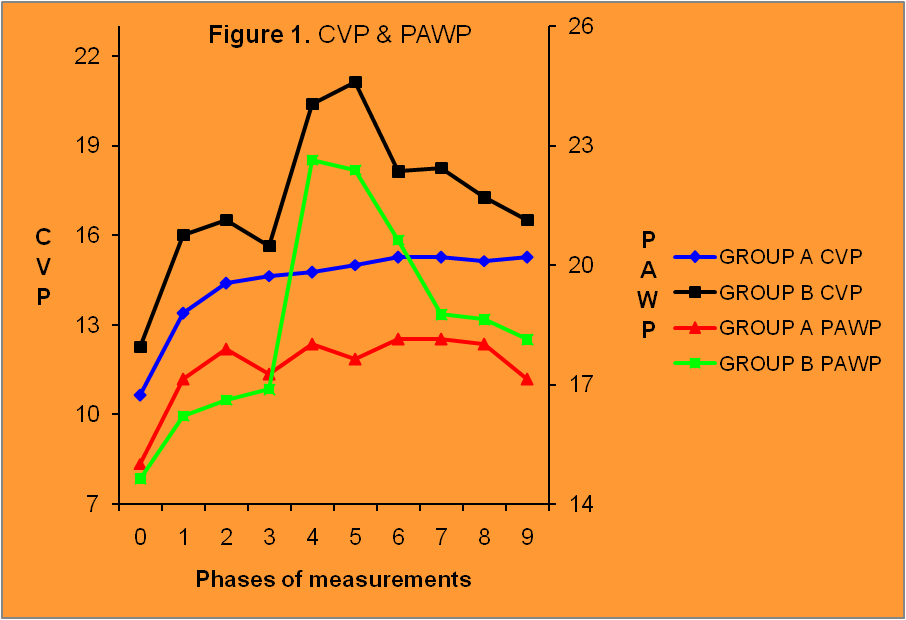
Increased IAP often coexists with sepsis in severely ill patients in the ICU, under mechanical ventilation and pharmaceutical support of the circulation with inotropes and vasoactive drugs. Both conditions have an unfavorable effect on the cardiovascular system. The purpose of this experimental study was to record the effect of increased intra-abdominal pressure on the cardiovascular system of pigs, with or without additional sepsis. Sixteen male pigs were randomly assigned in two groups A and B. In both groups, after induction to anesthesia and mechanical ventilation, the intra-abdominal pressure was increased to 25mmHg by helium insufflation in the peritoneal cavity, and that level of IAP was preserved until the end of the experiment. In Group A no other intervention apart from the increase in IAP was made, whereas in Group B, 60 minutes after the increase in IAP, 100μg/kg LPS were administered. Data were recorded after induction of anesthesia and initiation of mechanical ventilation (baseline measurement/measurement 0) and thereafter every 20 min after intra-abdominal pressure increase. The last measurement (measurement 9) was obtained immediately before release of pneumoperitoneum. Parameters measured or calculated included HR, BP(s,d,m), RVPs, PAP(s,d,m), PΑWP, CO, SV, SVR, PVR, SvO2, ETCO2. HR increased statistically significantly only in Group B, 60 minutes after the administration of LPS. BP (s, d, m) presented a significant change only in Group B, an initial increase immediately after LPS administration, followed by a decrease. CVP, RVPs and PAP (s, d, m) increased in both groups after IAP increase, whereas they presented an additional increase in Group B, after LPS administration. PΑWP changed only in Group B, after LPS administration. CO and SV were dramatically reduced in Group B, immediately after LPS administration, but gradually recovered their initial values until the end of the experiment. SVR changed only in Group B. They increased after LPS administration and then they gradually decreased. PVR increased dramatically after LPS administration and, despite gradual decrease they remained at high values until the end of the experiment. SvO2 decreased in Group B after LPS administration but gradually recovered its initial values. At the conditions of this particular experiment, the increase in intra-abdominal pressure was well tolerated by the laboratory animals. On the contrary, sepsis induction by LPS administration had an unfavorable effect on the cardiovascular system.
Continue readingAdequate adrenocortical function is essential to survive critical illness. The goal of this study was to determine whether eosinophilia could serve as a useful and early marker of adrenal insufficiency in critically ill patients with severe septic shock. During a 1-year period, we prospectively studied 294 ICU patients.16 patients (5.4% of ICU admissions) with eosinophilia more than 3% of the white blood cell count and septic shock unresponsive to adequate fluid and vasopressor therapy, were included. A high dose (250 mcg i.v) corticotropin stimulation test was performed. Eosinophilia (>3%) was diagnosed in 16 patients with vasopressor-unresponsive septic shock. Eosinophilia was present 1.9±0.9d (range 8-96h) before the onset of septic shock. 11/16 patients failed to respond to corticotropin stimulation test above the critical level of 9 mcg/dL rise and 2/16 had baseline cortisol concentration <10 mcg/dL. Baseline cortisol level, maximal cortisol increase post-corticotropin administration and Eosinophils count (%) were higher in survivors (p≤0.05). A hydrocortisone infusion (300mg/d) treatment resulted in haemodynamic improvement in 12 of 16 patients (75%). The 28-day mortality (following the onset of septic shock) was 43.7%. Relative eosinophilia may be considered as a useful and early bioassay for adrenocortical function assessment in critically ill patients with septic shock and assumed adrenocortical depression.
Continue reading
The case describes a late presentation of Infantile Hypertophic Pyloric Stenosis in a 5 year old boy, and its management.
Continue readingThymoma is one of the most common tumors of the anterior mediastinum in adults, arising from thymic epithelial cells. Complete surgical resection is the treatment of choice in myasthenic patients with thymoma. A 41 year-old male, with myasthenia gravis and thymoma was scheduled for thymectomy. Ten months ago, he developed pneumonia. He had a history of pneumonia (10 months ago) complicated by severe adult respiratory distress syndrome (ARDS), managed successfully with a short-term support with extracorporeal membrane lung assist device NovaLung. Long-term impairments in lung volume and diffusion capacity have been reported in adult respiratory distress syndrome survivors. Total intravenous anesthesia technique and especially muscle relaxants infusion require special attention in this group of patients. Our management strategies in this case included careful assessment of respiratory function, avoidance of premedication, use of short-acting anesthetic agents (propofol, remifentanil), use of suggamadex for reversal of rocuronium-induced neuromuscular block, and intraoperative monitoring with a train-of-four monitor and monitoring in an intensive care unit for 24 hours after surgery.
Continue reading
- Letter from the Editors
- Editorial
- Intracranial Hypertension. Pathophysiology-Symptoms-Monitoring.
- The clinical applications of body weight and endotracheal tube size prediction formulas in pediatric patients.
- The use of National Center of Emergency Care services by Refugees Camps in Northern Greece-6 month study.
- Anesthetic management of a patient with Myasthenia Gravis for abdominal surgery
- Pacemaker syndrome during temporary epicardiac pacing, after coronary artery bypass grafting: case report and review of the literature.
Dear colleagues,
The uninterrupted and continuous presence of the Greek e-journal of Perioperative Medicine in the scientific literature, over last 17 years, has been gradually increased and this is attributed to your help. On behalf of the editorial board, we would like to emphasize that our goal is ensure free-access, high quality, published articles in the area of medical press and we would like to thank you again for this continuous support. This is the third (3rd) issue of the year and our goal is to raise the issues from three to four per year. The participation of all of us with high quality publications will enable our journal to continue to be part of international libraries.
The present issue covers a wide area of interests. The review article of Schizodimos et al summarizes the major pathogenetic mechanisms, the clinical manifestations and the monitoring techniques of intracranial hypertension. They refer to 2016 Guidelines for the Management of Severe Traumatic Brain Injury, where a measured ICP above 22 mmHg is considered an indication for intervention. They also describe the major pathogenetic mechanisms of intracranial hypertension (venous occlusion, increased cerebral volume, increased blood volume, mass effect cerebral edema). Additionally, new techniques… Continue reading
The exchange of experiences and ideas among colleagues remains the key of scientific progress. In fact, knowledge is a sum of gathered experiences and education is nothing more than an attempt to pass all those knowledge to the next generation.
In rapid transforming scientific disciplines like medicine, the need for this exchange is essential. The latter can take different forms. Oral conversation may seem like the humble way of communication. Nevertheless, “conversation is a meeting of minds with different memories and habits. When minds meet, they don’t just exchange facts; they transform them, draw different implications from them, engage in new trains of thought. Conversation doesn’t just reshuffle the cards; it creates new cards”1-2.
Written documentation, in form of abstracts/posters/articles, is the formal way of medical communication. The ultimate goal: better clinical medicine and thus, better outcomes. Secondary goals: scientific research boost, professional progress, academic career evolution, financial remuneration, etc.
Continue reading
Increased intracranial pressure (ICP) is a serious final common pathway of a variety of neurologic injuries. Elevated ICP has consistently been associated with a poor outcome. It is a medical emergency requiring immediate intervention to prevent permanent damage to the brain. The Monro-Kellie doctrine states that the intracranial space is a fixed volume inside the skull. It describes the principle of homeostatic intracerebral volume regulation. The Monro-Kellie hypothesis and cerebral dynamics are important in order to understand the pathophysiology of intracranial hypertension. Venous occlusion, increased cerebral volume, increased blood volume, mass effect and cerebral edema are the major pathogenetic mechanisms of intracranial hypertension. The clinical manifestations of increased ICP are varied and unreliable. Headache, vomiting, disorientation, and lethargy are the main symptoms as well as symptoms and signs caused by cerebral herniation. ICP monitoring is widely used in clinical practice in order to improve patient outcome. It is especially useful as a robust predictor of cerebral perfusion, and can help to guide therapy and assess long‑term prognosis. Intraventricular catheters remain the gold standard for ICP monitoring, as they are the most reliable, accurate and cost‑effective, and allow therapeutic cerebrospinal fluid drainage. Intraparenchymal catheters are usually considered accurate, with the potential disadvantage that they measure localised pressure, which may not be reflective of global ICP. Furthermore, non‑invasive methods of ICP monitoring, such as transcranial Doppler, optic nerve sheath diameter, etc., have emerged as promising techniques for screening patients with raised ICP in settings where invasive techniques are either not feasible (patients with severe coagulopathy) or not available (setups without access to a neurosurgeon).
Continue readingThere are various formulas and algorithms for the prediction of body weight and appropriate ETT size, in pediatric patients. Body Weight estimation is of paramount importance in pediatrics, especially in emergencies. Predicting the appropriate size of the endotracheal tube saves time, money and reduces complications. The goal of this study was to evaluate the validity of two commonly used formulas for predicting the body weight and the size of the appropriate endotracheal tube, both based on age.353 consecutive pediatric surgical patients aged 2 to 12 years, who required general anesthesia and oral endotracheal intubation were included in this study. Patients were stratified according to their age in two groups: group 2-5 (79 children, 2 to 5 years) and group 6-12 (274 children, 6 to 12 years). At the end of surgery an anesthesiologist, who was not involved in the perioperative treatment, recorded the demographic data and also the size and type of the endotracheal tube used. The prediction of Body Weight (BW) was made according to the following formula: 2-5 y.o.: Weight (kg) = (2 x age in years) + 8 and 6-12 y.o.: Weight (kg) = (3 x age in years) + 7. The formula for calculating the size (size= internal diameter=I.D.) of the endotracheal tube (ETT) was: I.D. for cuffed ETT (mm) = (age / 4) + 3.5 and I.D. for uncuffed ETT (mm) = (age / 4) + 4. For all statistical tests p value <0.05 was considered as statistically significant. In all patients as sum and in both age groups, the predicted body weight was significantly (p<0.05) lower than the actual (measured) weight. In group 2-5y.o, 74.7% of patients received cuffed ETT In group 6-12y.o. 100% of patients received cuffed ETT. In group 2-5 y.o, all patients showed a significantly (p<0.05) lower predicted internal diameter of the ETT, either cuffed or uncuffed, compared to ETT ultimately used. In group 6 -12y.o, there was no statistically significant difference between the predicted and the actually used ETT size. The prediction of body weight in children, by the use of the particular formula, led to underestimation. In children aged 2 to 5 years, the application of the inner diameter calculation of the ETT formula also underestimated the appropriate ETT size. It seems that the traditional age-based formulas often fail to predict the correct ETT size in smaller children which probably does not seem to apply to older children.
Continue reading


